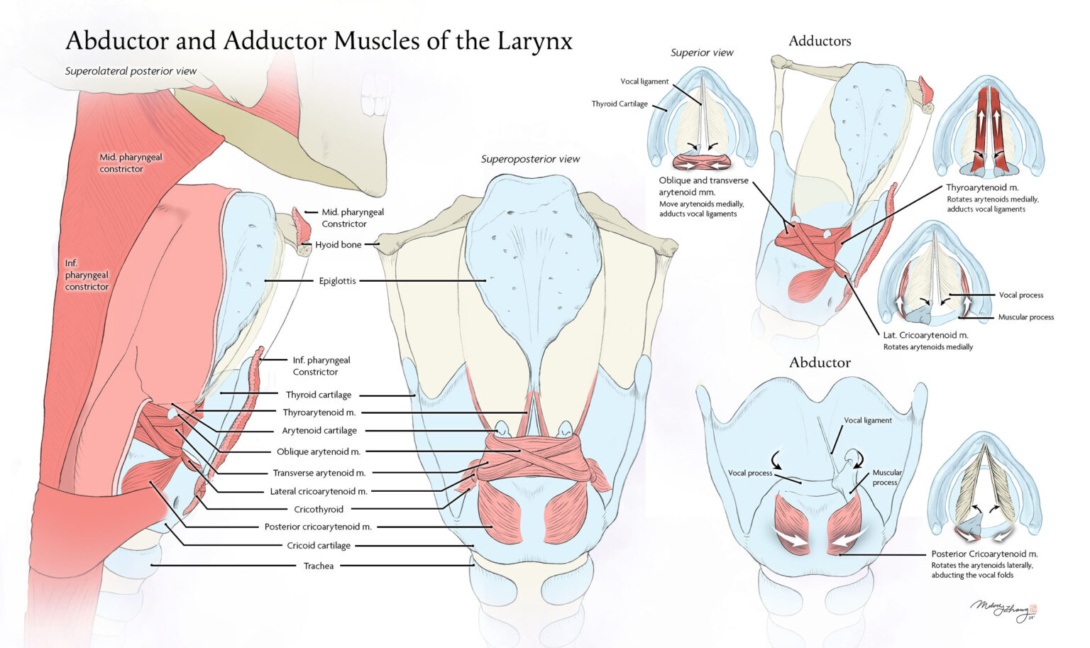
This anatomical illustration highlights the adductor and abductor muscles of the larynx in oblique and superoposterior views. It was created to help medical students understand larynx muscle contractions (yellow arrows) and the correlating arytenoid cartilage movements (black arrows) that allow vocal ligaments to adduct and abduct.
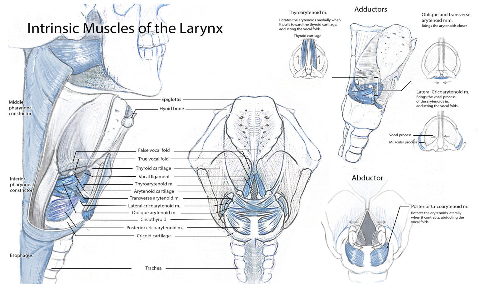
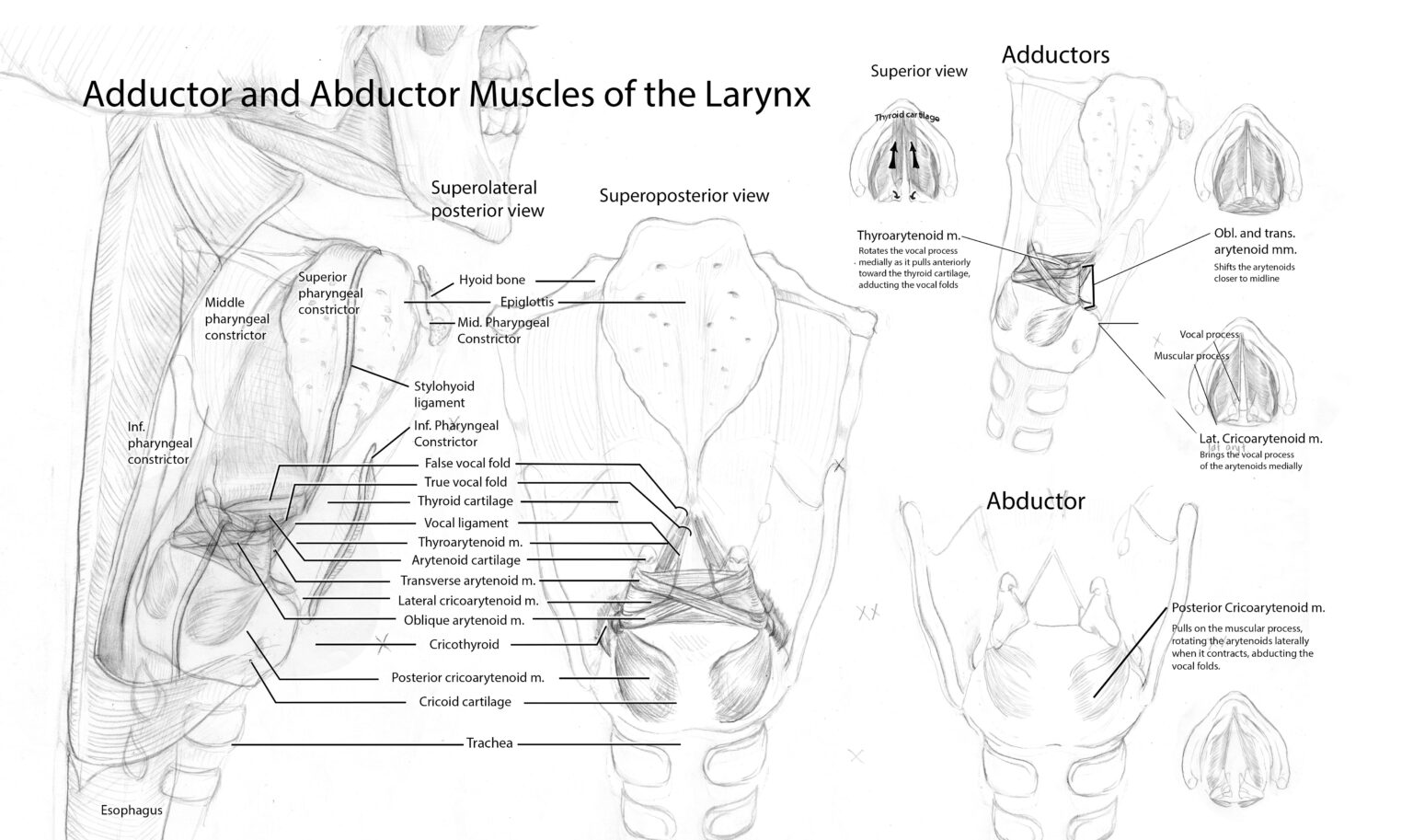
The image to the left depicts a full view of the larynx, with an inferior skull and pharyngeal constrictors cut to show the location of the laryngeal structures. The mucosa is cut to see the cartilage beneath, and the muscles are ghosted to reveal arytenoids. The central image is a summary view of laryngeal musculature with shared labels used to connect and contextualize shared structures. The central top right figure utilizes an oblique view to depict adductor muscles, which are further clarified with insets shown superiorly. A superoposterior view in the bottom right figure highlights the movement of the abductor muscle and includes another superior inset.