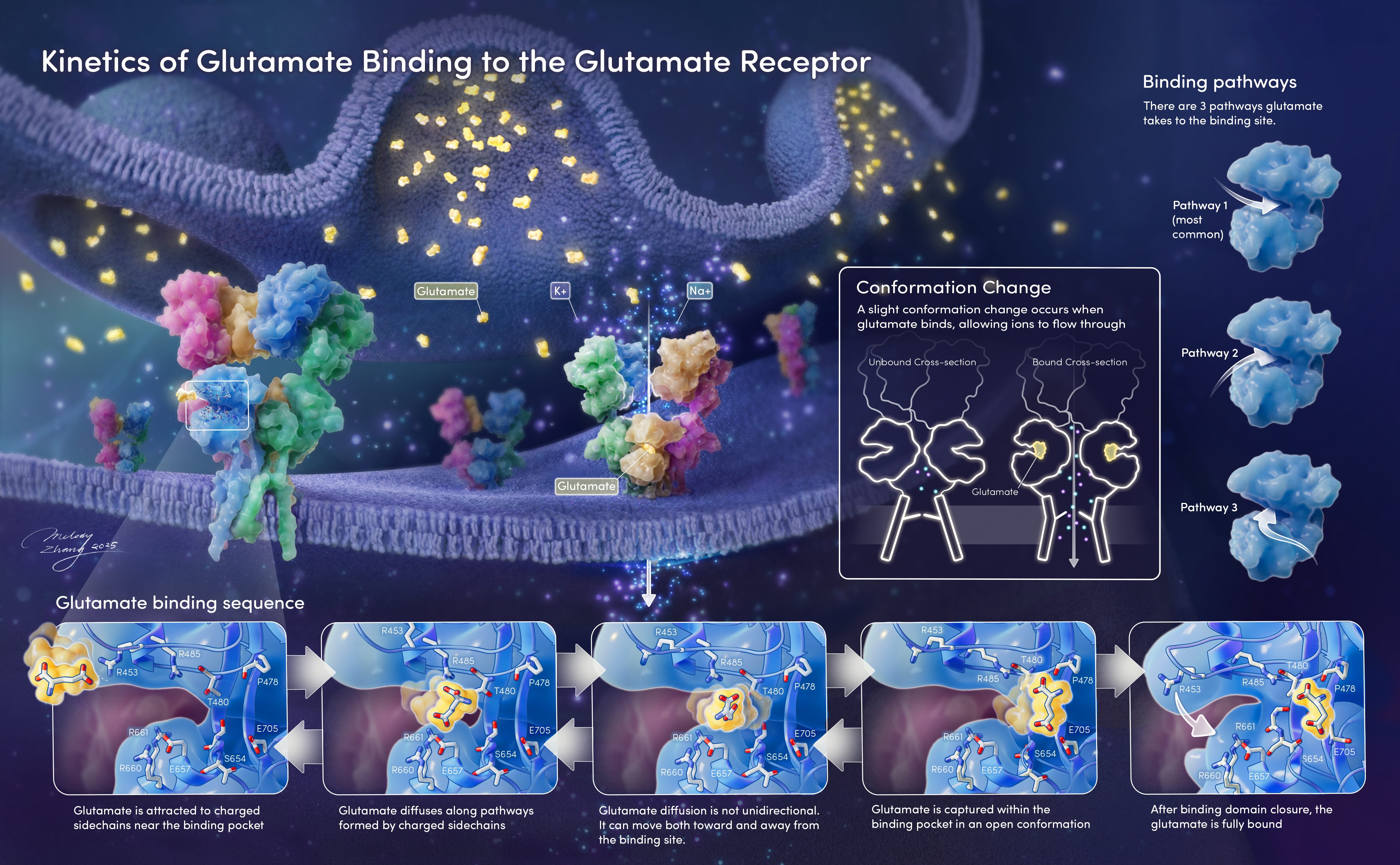
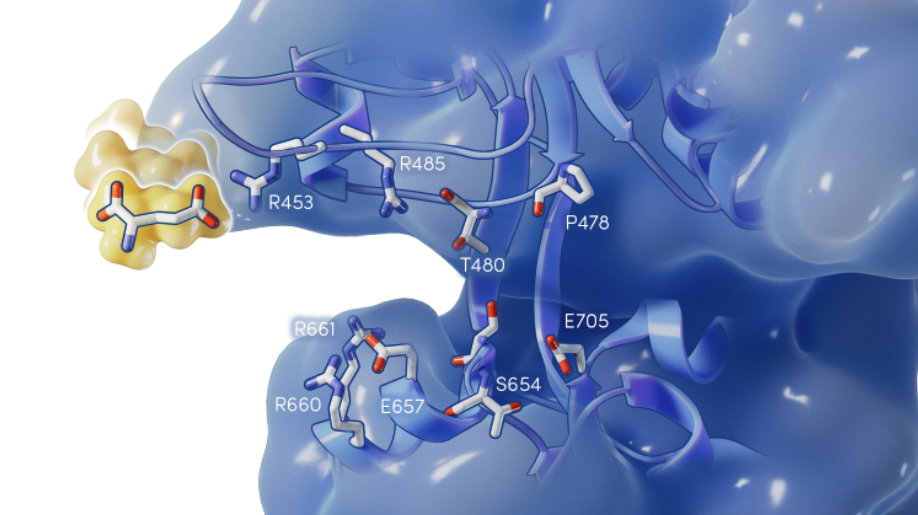
This molecular illustration highlights the binding sequence of glutamate when it binds to a glutamate receptor, showcasing a dynamic zoom in on the synaptic cleft. The client requested a 5 step sequence demonstrating the movement of the receptor side chains, as well as a diagrammatic explanation of the conformation change. The intended flow is as follows: viewers will be guided by the release of glutamate molecules from the synaptic vesicle. The main image features a glutamate approaching a receptor from the left, and viewers may choose to follow the binding sequence from the zoom in or veer right and inspect the conformation change that occurs when the glutamate is bound. At the very right, the client requested that 3 common pathways glutamate takes to bind are shown.
Process
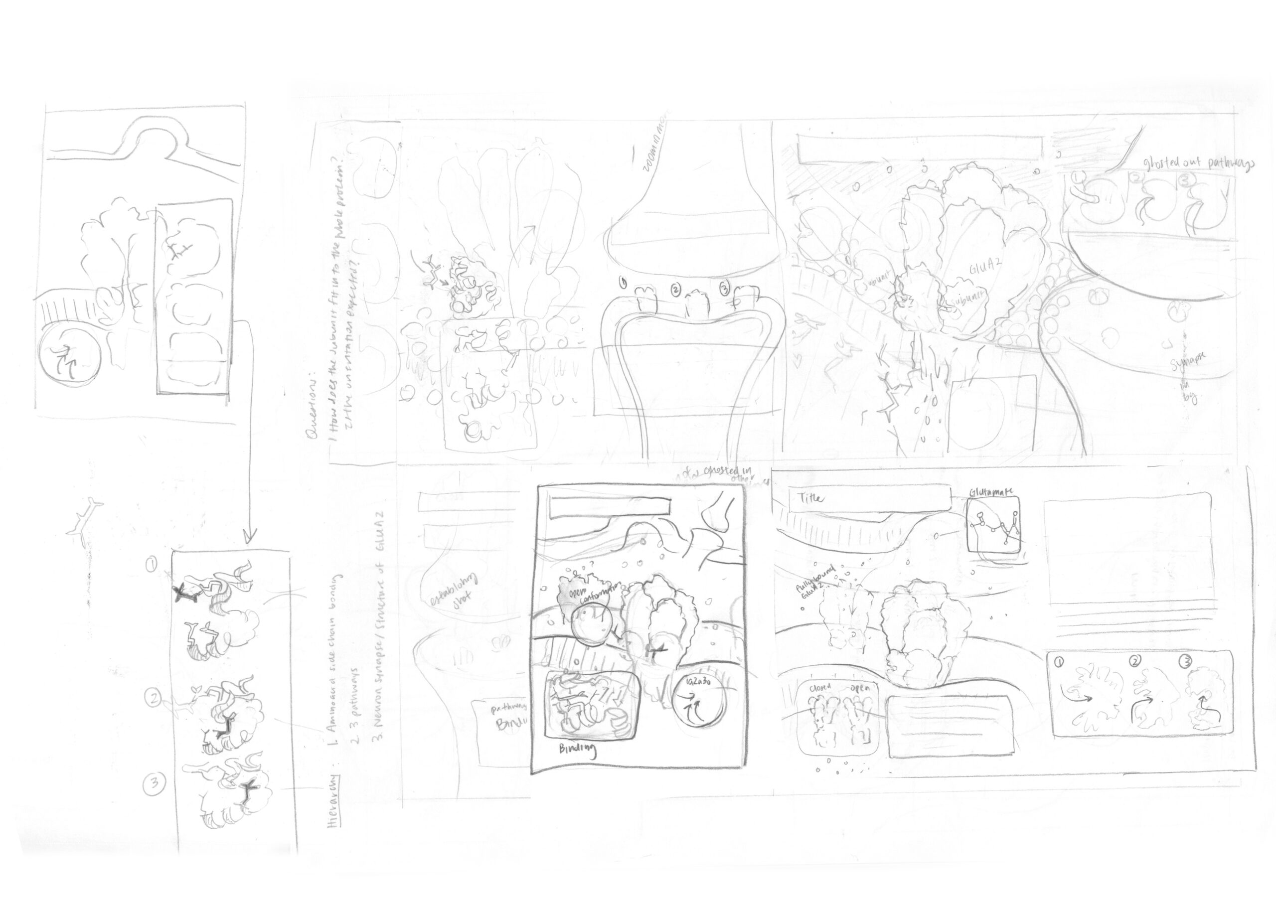
Thumbnails
The process began with a series of thumbnails. After reviewing the thumbnails with the clients, the thumbnail or combination of thumbnails with the greatest teaching value are chosen.
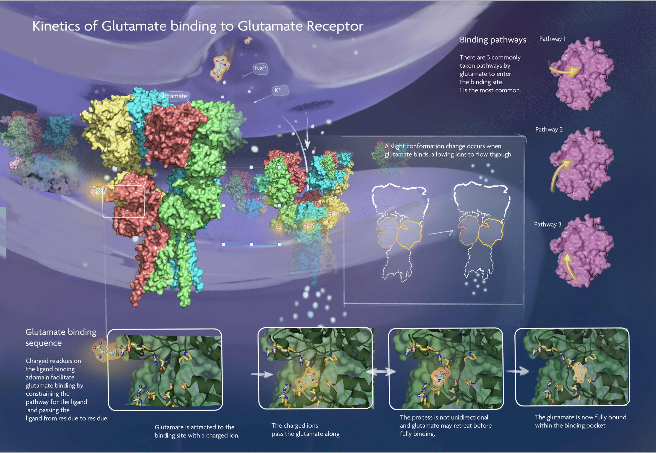
The initial assets are found using Pymol and quickly placed into the photoshop layout to mock up the rough layout. Initial colors are direct from Pymol and will be adjusted in Cinema 4D.
Pymol assets and rough sketch
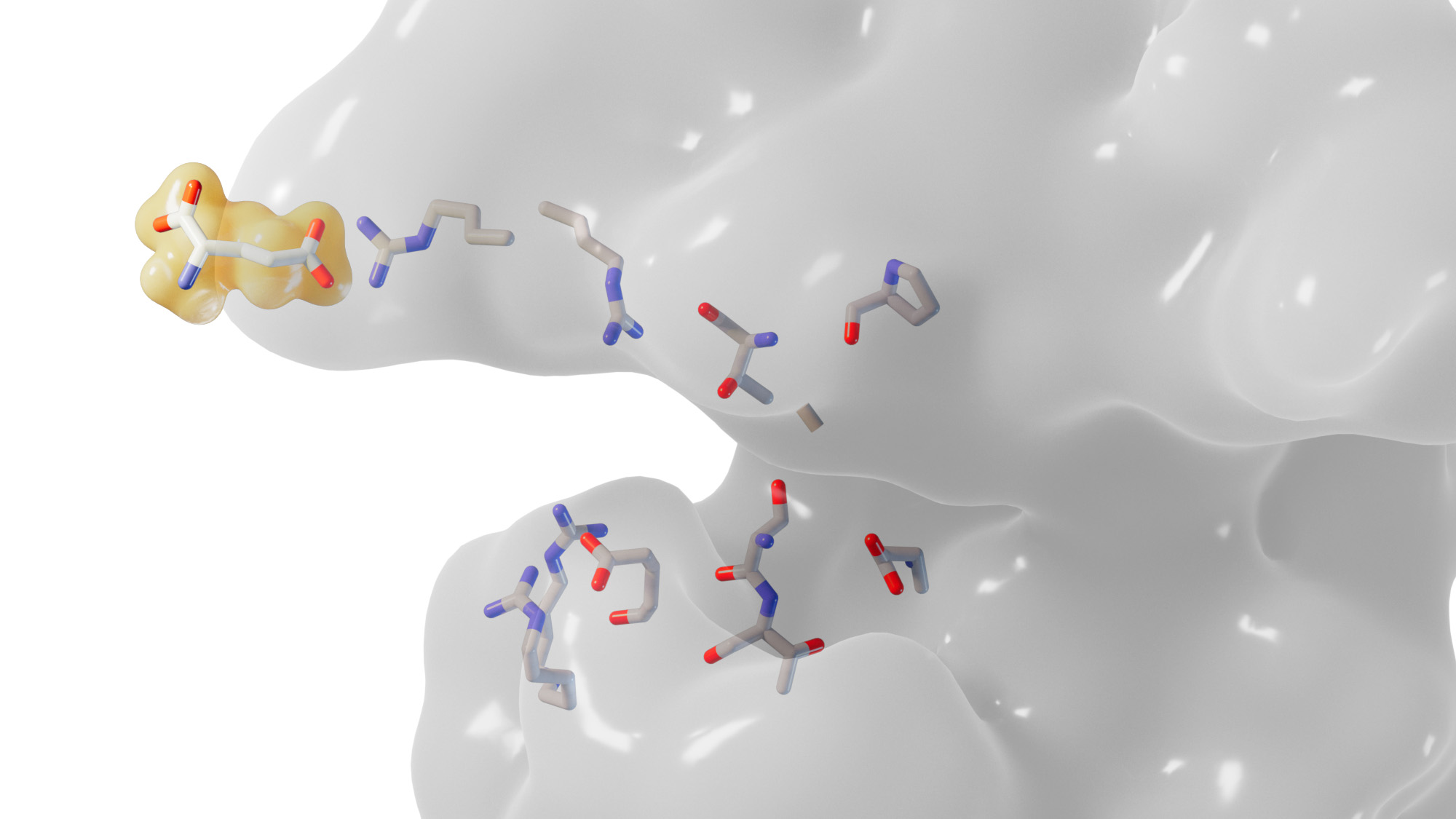
Camera View and Final
Side chains and glutamate assets
Glutamate receptor asset
Through iterative review and feedback, the assets were rendered individually from Cinema 4D, with additional ambient occlusion and subsurface scattering passes made to allow for maximum adjustment in Photoshop. The final piece is a composition of assets from Cinema 4D, diagrams from Illustrator, and additional line, text, and masking elements from Photoshop.