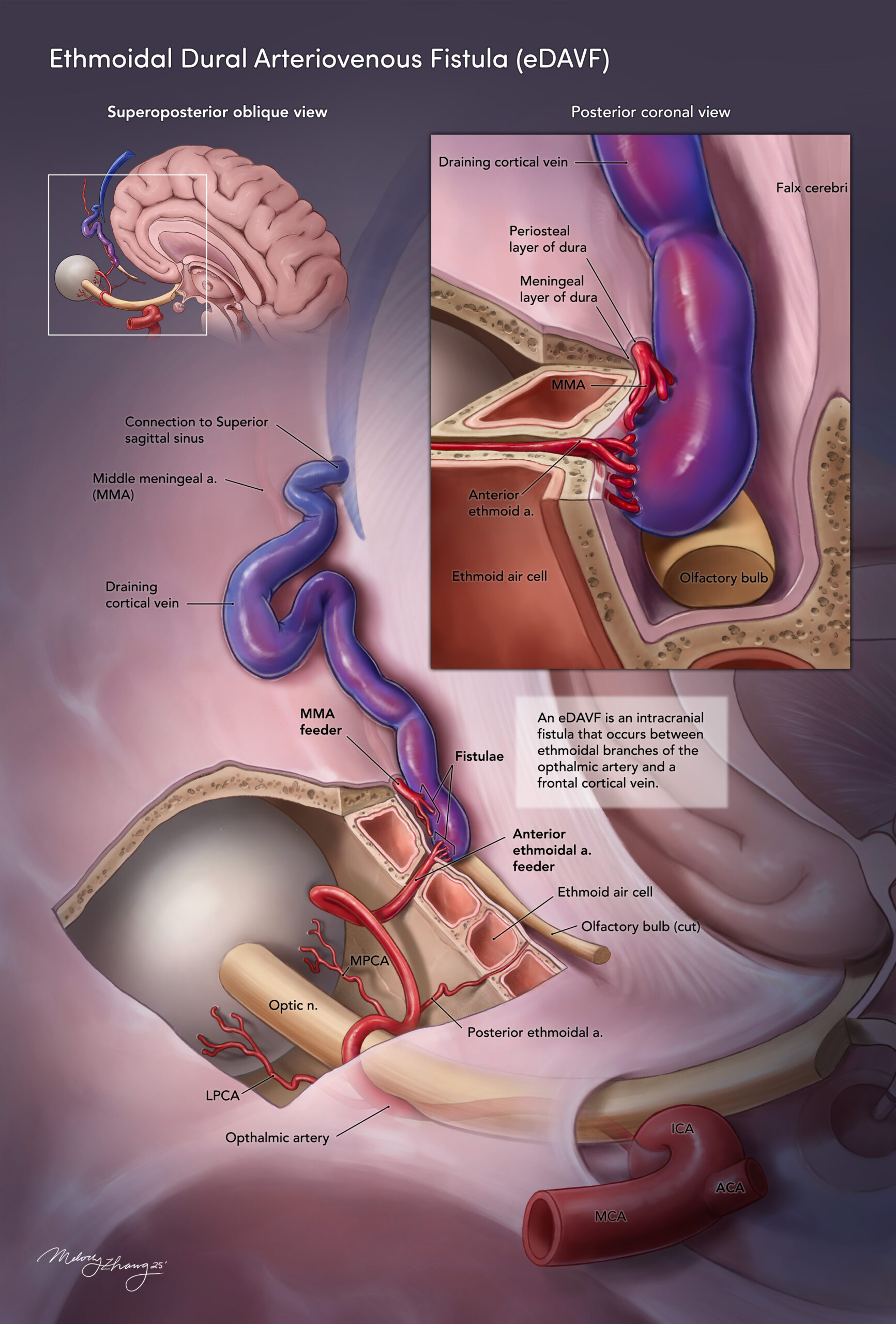
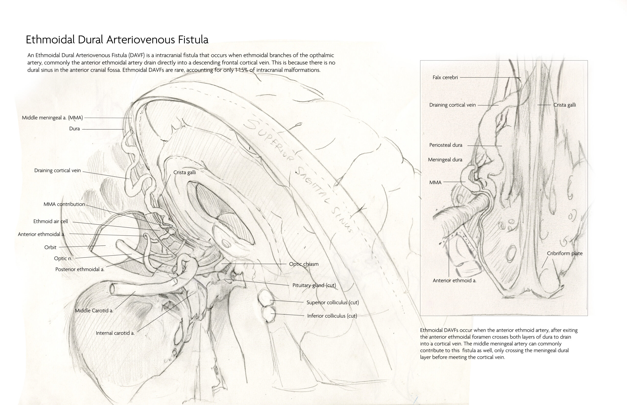
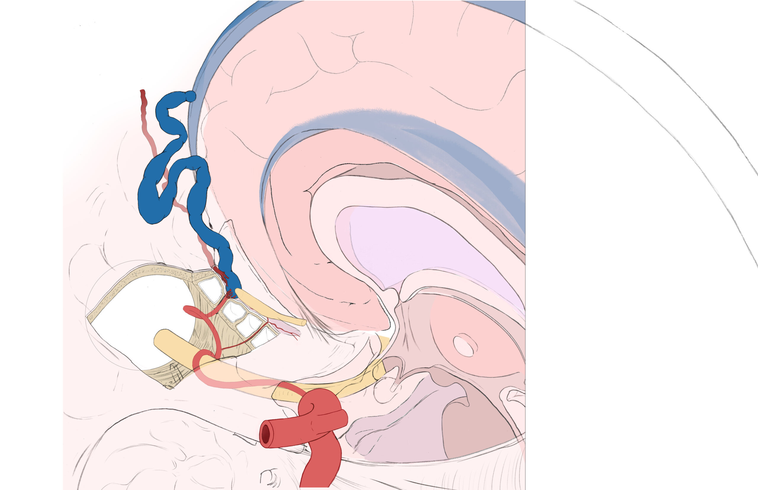
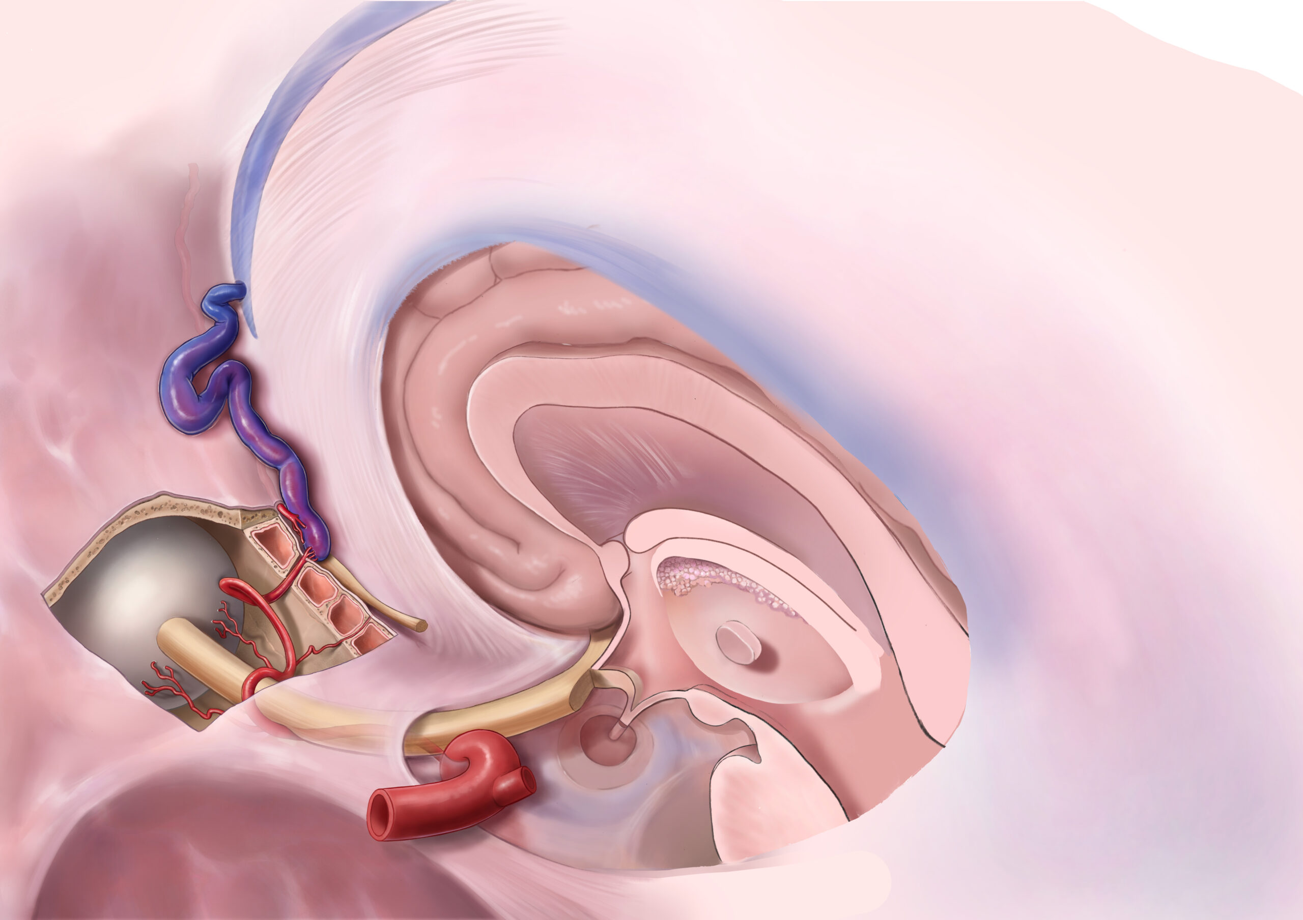
This neuroanatomical plate was created for Dr. Risheng Xu, a neurosurgeon at the Johns Hopkins Hospital. The focal image is within the skull, with the large arteriovenous fistula located at the cribriform plate. Half of the brain is removed to more clearly show the relation between structures. An inset on the top left is used to orient the viewer and the inset on the top right gives a close up, frontal view of the fistula.